 Laboratory of Anatomy, Biomechanics and Organogenesis
Soft tissue reconstruction by digital stereophotogrammetry
Soft tissue (muscles, ligaments) 3D morphology reconstruction, including extraction of muscle and tendon fibre topology and attachments are developed as a software pipline in this research. It contains 3D reconstruction of dissected soft tissues as alternative to high resolution MRI that shows some limitations when whole specimen body has to be scanned.
This research developed a method using stereophotogrammetry on the basis of digital cameras allowing soft tissue data collection and maintaining anatomically-correct results. The method was based on an image recognition toolbox and was validated by processing simultaneous poses of calibrated clusters made of coloured balls and pins attached to a 3D digitizer (Platinum FaroArmc 4ft., accuracy = 0.013mm). Three pre-calibrated digital cameras (Olympus SP-500UZ, 6 mega pixels) allowed reconstruction accuracy about 1 mm for the captured volume (0.5x0.5x1.0m3).
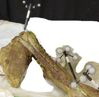
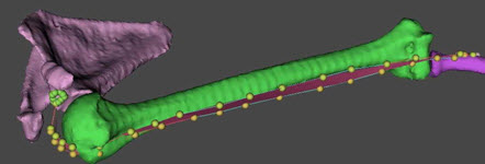
Prior to dissection, a specimen has been fully CT-scanned to obtain bone 3D models. During specimen dissection, pins with colored heads (COHs) were inserted in muscles and ligaments to characterize muscle and ligament fibre path, musculo-tendinous junctions, origins and insertions (Top image). CHO data registration to the 3D bone models occurred using technical frames including reflective markers and aluminium balls inserted into the bones for better accuracy (TF) rigidly attached to the bones. Once registered, CHO information was reconstructed in 3D together with the bone models (bottom image). Muscle and tendon fiber length and pennation angles were evaluated after piece-wise linear approximation of the reconstructed points. The method has been applied on an entire human body.
Top image: TFs (right scapula, humerus, radius) and colored pins captured by digital camera for biceps brachii (caput longus) muscle. Bottom image: Final reconstruction and visualization in lhpFusionBox software. |
|
Need more information? Please contact ![]() !
!